

The skin is a good model for the study of ageing because of its accessibility and also because its cellular elements– keratinocytes, melanocytes, and fibroblasts, among others– can be easily grown in cell cultures and used in cellular and molecular research.
Some scientific research carried out using these methods indicates that skin senescence follows the general principles of aging that depend on the body‘s biological clock. However so-called “crono-aging” is also strongly influ- enced by external environmental factors, including solar ra- diation, which aggravates the senescence of skin regions exposed to sunlight, thus called photo-ageing.
These two processes are characterised by different clinical aspects influencing two different kinds of skin wrinkledness. Crono-ageing, a fine wrinkledness, is accompanied by skin hypotrophy, progressive loss of the subcutaneous fat, and subsequent drooping. In photo-ageing, visible symptoms include deep wrinkles and skin folds, skin thickening, pigmentation disorders (eg, lentigo solari, or liver spots), and telangiectasia.
Both chrono-ageing and photo-ageing can occur in skin areas damaged by sunlight, but which condition will predominate depends on the absorbed cumulative radiation intensity as well as genetic and racial factors.
Crono-ageing
From a histological point of view, crono-ageing is characterised by a flattening of the dermal and skin interface with reduction of the corium papilla (dermal papilla). The epidermis becomes thinner and flattened, and cells of the basal and prickle layers appear heterogeneous and less well organised. In affected regions, cells display changes in size, shape, and chromophilia and gradually lose their specific polarity.
Sometimes some “dark” cells are seen, which appear to be dyskeratotic and necrotic. Over time, the corium gets increasingly thinner with an apparent increased density of collagen fibers. This collagen increase is thought to be caused by both the reduction of the spaces between the fiber bundles – often occupied by highly hydrophilic glycosaminoglycans (in particular, hyaluronic acid) – and the reduction of elastic fibers, implying that the connective tissue is becoming more compact due to a low concentration of water. Moreover, the collagen fibers become harder and the skin becomes less elastic, probably due to changes in intermolecular and intramolecular bonds.
Elastic fibers start to disintegrate, being transformed into shorter fibrils with visible cysts and lacunas. Hair follicles shrink, and sebaceous and sudoriferous glands hypotrophy. Circulation becomes compromised, characterised by reduction in the size and quantity of capillaries as well as increased basal membrane thickness.
Photo-ageing
Epidermis subjected to photo-ageing appears thickened (acanthosis), with occasional cellular dysplasia and variable cellular atypia. At the dermal level, the elastic fibers can become thickened and degenerate into a mass of amorphous matter (solar elastosis). Conversely, collagen fibers are reduced in volume, with an attendant increase in soluble collagen levels.
Proteoglycans, which are quantitatively reduced in intrin- sic aging, increase in chronic actinic injury. Vessels become dilated and tortuous; mastocytes become partially degranulated, with possible release of proteases, which are partly responsible for degenerative phenomena. An increase in inflammatory cells is seen in photo-ageing, whereas the intrinsic ageing process is characterised by a decrease in the overall number of cells.
Skin Electroporation
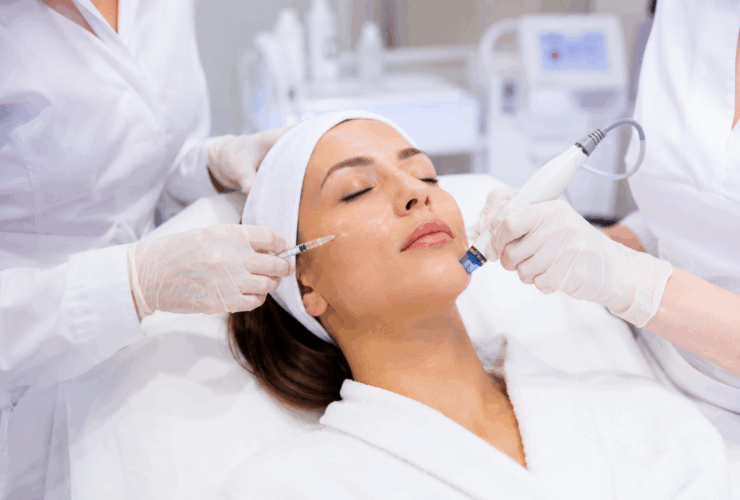
This is a non-invasive technique based on the transitory increase of the skin permeability caused by a particular electromagnetic wave, generated by an high-tech electronic device, developed by the Microlab Biomedical labs. Skin electroporation occurs when new ducts for water-soluble substances are created in an epithelial cell by an appropriate en- ergy administration.
Such alteration makes the cell membrane more permeable for a wide range of hydrophilic molecules which could not enter the cell otherwise. Once formed, these ducts stay open for only a short only time. The permeability increase on the target tissue highly favors the absorption in the deepest skin layers of those macromolecular structures such as collagen, elastin, and hyaluronic acid, which are essential for the reduction of actinic injuries.
This technique is easy and totally harmless, enabling the transfer into the skin of those substances the doctor considers suitable to regenerate, tone up, and smooth the patient’s skin.
Results
At the first application results are surprisingly clear, even if the areas mostly subject to micry showed a loss of the optimal calls for on the third or fourth day. For this reason the protocol foresees 2 weekly applications for 6 weeks and a further weekly application for the next 6 weeks and, if necessary, a monthly follow-up visit.
Table
Assessment of treatment results. The most improved parameters were moisture and tonicity, as expected.
- Assessments by Individual Patients
- Assessments by Videocapillaroscopy by Optic Probe
Discussion
The results of this clinical study show a 100% medical improvement of tonicity and moisture and a 70% improvement of the other parameters.
The advantage of such a technique is the absence of side effects and few contraindications (excluding pacemaker patients, pregnant women, and patients with metallic implants).
Patients’ compliance has been total since treatments have been well tolerated, with the exception of a metallic taste in the mouth. To date the obtained results are better than any other modern medical cosmetic technique, even with the same administration of biointeractive hyaluronic acid, since with electrotransmission it can be more homogeneously spread, thus giving better results.
Conclusion
The effectiveness, the absence of side effects, and the high comfort level of patients noted in this study show that the use of transdermal electroporation for the transmission of hyaluronic acid can be used to replace traditional invasive needle techniques used in skin rejuvenation for particularly aged skin, and it is also ideal for patients not wishing to be subject to invasive treatments.
Claudio Amitrano, MD Member, European Society of Cosmetic & Dermatological Surgery 80126 Napoli (Its Via Cinthia, Parco S. Paolo is.
Tel/Fax 011 390 817676183